标题: Low expression of PDK1 inhibits renal cell carcinoma cell proliferation, migration, invasion and epithelial mesenchymal transition through inhibition of the PI3K-PDK1-Akt pathway日期:2018年11月20日
单位与作者:
南昌大学江西医学院Wei-Min Zhou、Gong-Xian Wang(通讯作者 音译 王共先)
江西省肿瘤医院泌尿外科Xin-Hua Tu(通讯作者 音译 涂新华)
期刊:Cellular signalling
#1 我对这些 WB 上没有背景以及条带保持完全线性并在凝胶向上移动时保持蛋白质团形状的方式很感兴趣。
Figs 5D,H.I am intrigued by the absence of background on these WBs, and the way the bands remain perfectly linear and retain the shape of the protein blobs as they move up the gel. Is it possible for the authors to upload the original scans of the gels?
#2 图10A与Mao et al (2018)的8A出现重复。Fig 10A. "Si-PDK1 inhibits cell invasion of RCC cells. A, the invasive A498 cells in each group".Orange box for comparison with Fig 8A of Mao et al (2018).
#3 图10A与Fan et al (2020)的6a出现重复。[below] Fig 10A again. "Si-PDK1 inhibits cell invasion of RCC cells. A, the invasive A498 cells in each group".
[above] Fig 6a from Fan et al (2020). "Cells in the basolateral chamber were observed under an inverted microscope".
#4 图10A与Fan et al (2020)的6a出现重复。
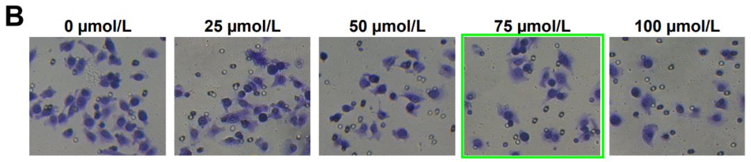
Another sighting! Fig 2B from "Mifepristone inhibits proliferation, migration and invasion of HUUA cells and promotes its apoptosis by regulation of FAK and PI3K/AKT signaling pathway" (Sang et al 2018).
参考信息:
https://pubpeer.com/publications/925EAD41B9C7AA6D0D78BEF8BE739B#4
https://www.sciencedirect.com/science/article/abs/pii/S0898656818302869?via%3Dihub